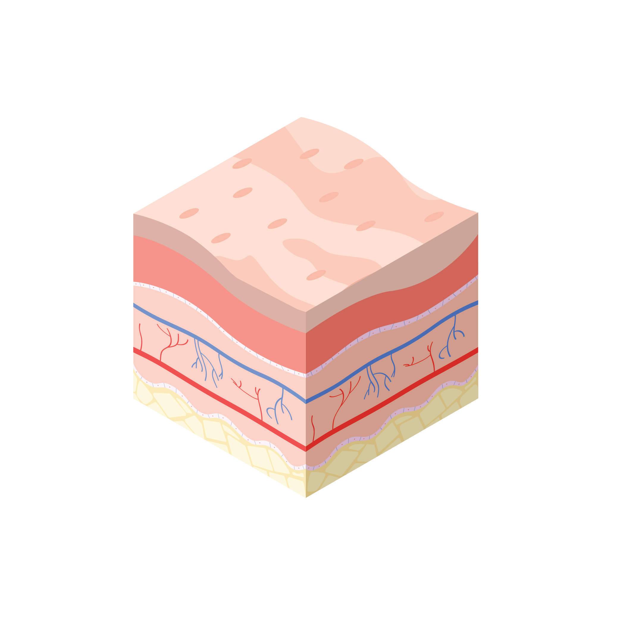
What is an Epithelialising Wound?
Epithelialisation is the final stage of wound healing and is pink/white in colour. It is the final stage of wound healing and only occurs on top of healthy granulation tissue. During epithelialisation, growth factors stimulate the proliferation of new cells and their migration across the wound. Once they cover the wound surface and meet, they stop dividing - known as contact inhibition. The wound surface is covered by new epithelium and the barrier function of the skin is now restored.
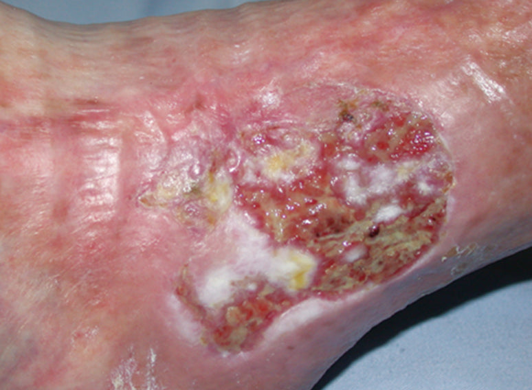
Treatment Aims
Care is needed when cleansing the wound and when removing dressings. Strong adhesives should be avoided to prevent skin stripping and shearing damage. Treatment of epithelialising wounds requires the creation of a warm, moist environment.¹ It is important not to allow the wound to dry out, as the epithelial cells will have to burrow deeper underneath the scab to close the wound, and this will prolong healing and result in a less cosmetically acceptable appearance. ²
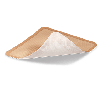
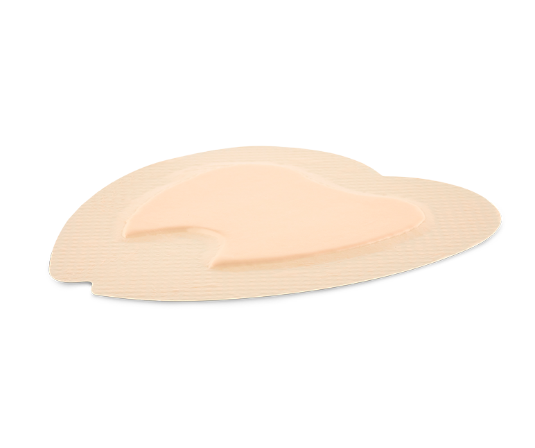
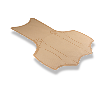
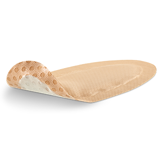
Products that treat Epithelialising Wounds

Learn More
With the Education HubWant to learn more about a range of different wound types and how they can be treated? Visit our free Education Hub.
Wound Types
Select a wound type below to discover more...
-
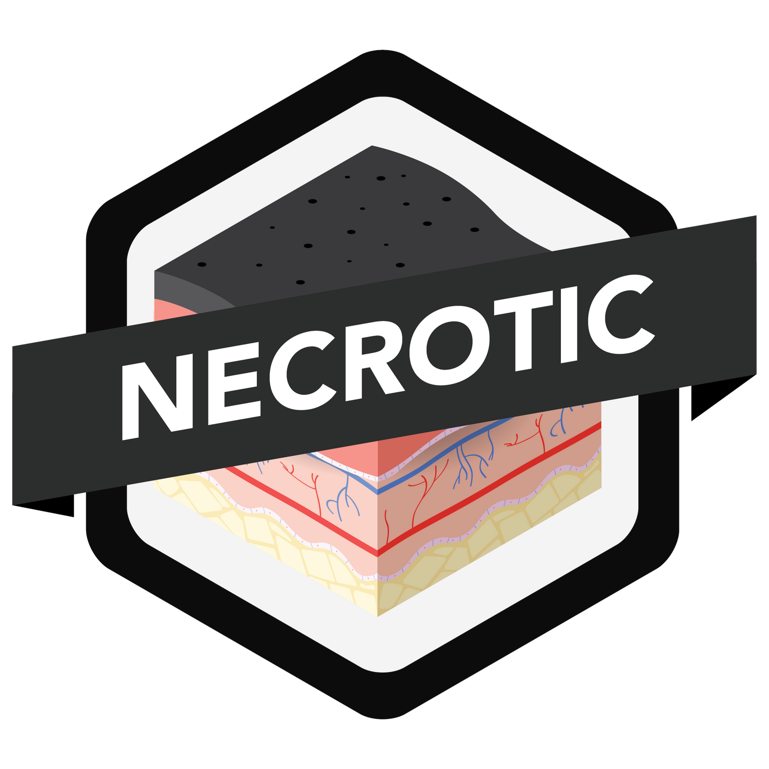
Necrotic tissue is dead or devitalised non-viable tissue which impedes wound healing.
-

Slough refers to the yellow/white material in the wound bed.
-

Granulation describes the appearance of the red, bumpy tissue in the wound bed as the wound heals.
-
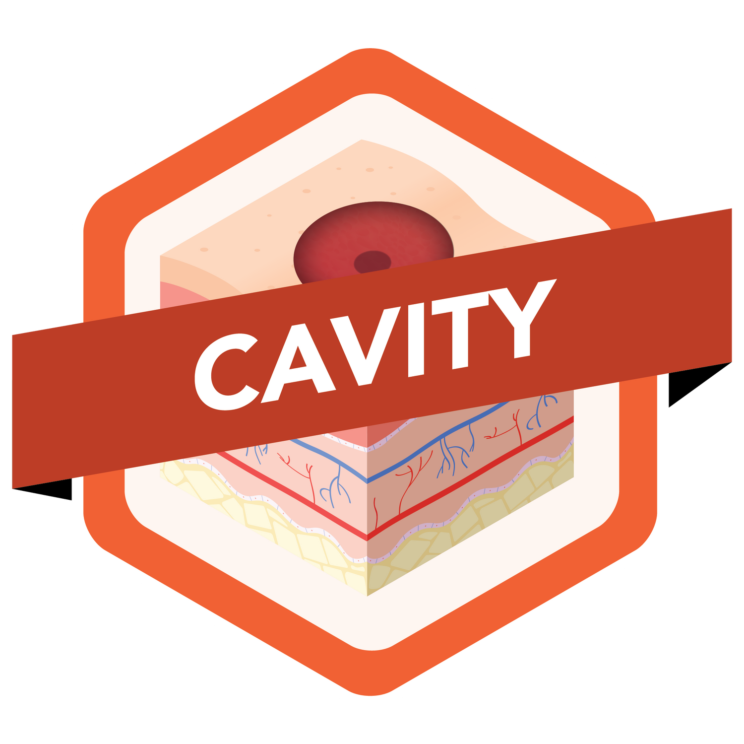
Cavity wounds can be defined as a wound that extends beneath the dermis.
-

A fungating wound develops when cancer that is growing under the skin breaks through the skin and creates a wound.
-

A scar may appear flat, lumpy, sunken, or coloured.
-
Infection can develop in any type of wound and is usually accompanied by pain, inflammation and swelling.
-
Return to the Wound Guide Information Page
References
1. Winter GD (1962) Formation of the scab and the rate of epithelialisation of superficial wounds in the skin of the domestic pig. Nature 193: 293–4
2. Schultz GS, Sibbald RG, Falanga V et al (2003) Wound bed preparation. Wound Repair Regen 11(Suppl 1): S1–28


